
In 2019, scientists found clues to how the living coelacanth’s hinged skull grows and why its brain stays tiny, shedding light on skull evolution |

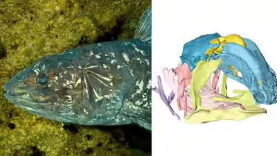

In 2019, scientists unveiled the first observations of how the skull and brain develop in the living coelacanth, Latimeria chalumnae, a deep-sea fish once thought extinct for seventy million years. A team working with the National Museum of Natural History in Paris scanned rare museum specimens without damaging them and published the results in the journal Nature. The findings shed light on a hinged braincase and a surprisingly small brain. The coelacanth’s braincase is split by a joint, and its brain fills only one per cent of the cavity, a mismatch unequalled among living vertebrates. This opened new avenues for research.
A coelacanth thought extinct for seventy million years was caught alive in 1938
The European Synchrotron explains that the coelacanth is a marine fish closely related to tetrapods, the four-limbed vertebrates that include amphibians, mammals and reptiles. Scientists believed coelacanths had died out seventy million years ago, until a South African fisherman accidentally caught a living one in 1938.Eighty years after that discovery, Latimeria still matters to researchers who want to understand where tetrapods came from and how their closest fossil relatives, the lobe-finned fishes, evolved. The study ‘Neurocranial development of the coelacanth and the evolution of the sarcopterygian head’ was led by Hugo Dutel, a research associate in palaeobiology at the University of Bristol, and was published in Nature in April 2019.
A hinged braincase and a tiny brain, the puzzling features of the living coelacanth
According to the report, one of the coelacanth’s most unusual features is its hinged braincase. Among the other animals that share it are many fossil lobe-finned fishes from the Devonian period, roughly 410 to 360 million years ago. A joint called the intracranial joint splits the coelacanth’s braincase completely into a front part and a back part.The brain itself sits far to the rear of the skull and fills only one per cent of the cavity that houses it. The article says this mismatch is unequalled among living vertebrates. How the skull grows and why the brain stays so small had puzzled scientists for years, so the team examined specimens at different stages of development from several public natural history collections.

Coelacanths live in the deep ocean and are a critically endangered species (Image: ESRF)
Scanning a five-centimetre fetus with X-rays to build three-dimensional models
Adult coelacanths are well represented in museums, but fetuses are extremely rare. The researchers therefore used advanced imaging to see inside the specimens without harming them. The European Synchrotron reports that they digitised a fetus only five centimetres long, the earliest stage available for Latimeria, using synchrotron X-ray microtomography on a beamline called ID19.The team combined those data with magnetic resonance imaging and micro-computed tomography scans to build detailed three-dimensional models. The models showed how the skull, the brain and the notochord, a tube running below the brain and spinal cord in early life, change from fetus to adult. The researchers also compared their observations with what is known about skull formation in other vertebrates.
An enlarging notochord may shape the braincase and the brain’s relative size
In most vertebrates, the report explains, the vertebral column replaces the notochord early in embryonic development. In Latimeria, the notochord instead expands considerably. The authors suggest this enlargement likely influences how the braincase is patterned and might underpin the formation of the intracranial joint. It may also affect the brain, whose relative size drops dramatically during development.
Source link